Imaging fractures and porosity in volcanic rocks
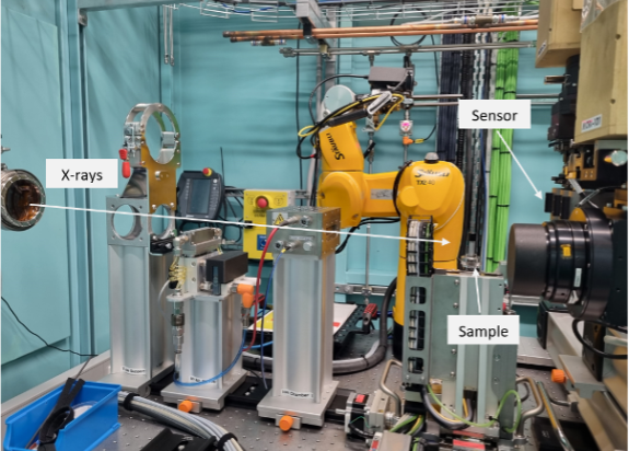
Our Massey University team members, Maia Kidd and Gabor Kereszturi, recently visited at the Australian Synchrotron in Melbourne. Their goal is to use Micro Computed Tomography (MCT) to image the macro- and microfracture networks in volcanic rock samples.
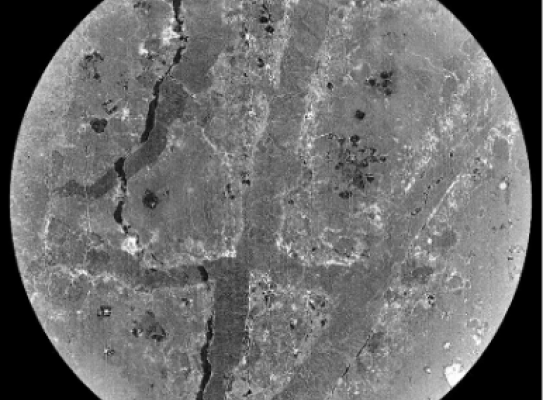
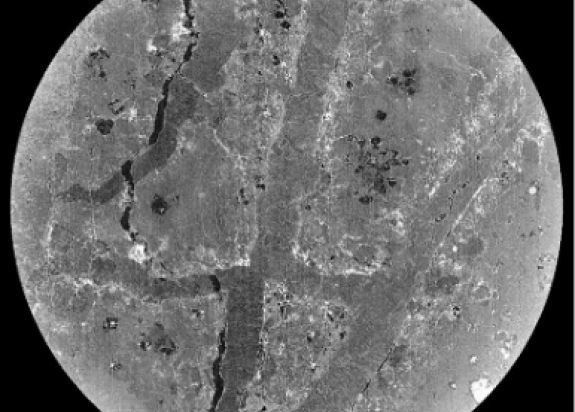
MCT is a non-destructive technique, which uses monochromatic, pink, and white X-ray beams, to provide detailed three-dimensional (3D) architecture at high spatial resolution (down to 6.5 microns).
The team collected images of over 40 rock samples from Whakaari (White Island), Ruapehu and Tongariro volcanoes, each with varying hydrothermal alteration.
The images collected are the first stage in a two-stage study. The synchrotron data is being processed to extract porosity and structural information, which will be used as pre-deformation baseline data for those rock samples.
In early 2024, the samples will be subjected to deformation experiments to measure triaxial rock strength. Then, the synchrotron scans will be repeated at the MCT beamline to highlight the change in the samples’ macro- and microfracture network. The pre- and post-deformation MCT data will allow the team to quantify the newly formed fractures and the changing porosity due to mechanical stressing.
This is important for understanding how hydrothermal alteration impacts rock deformation at the micro scale, and the potential impacts on slope stability and eruption dynamics.

Gabor Kereszturi
Earth Scientist

Maia Kidd
PhD Student